Command Palette
Search for a command to run...
From Membrane Segmentation to Particle Picking, Deep Learning Framework MemBrain v2 Streamlines Cryo-EM Analysis Pipeline, Significantly Enhancing Processing Speed and Throughput
Cryo-electron tomography (cryo-ET) is a powerful imaging technique capable of three-dimensional visualization of molecular environments within native cells at sub-nanometer resolution, offering unique insights into organelles, membrane structures, and macromolecular complexes. However, the biological complexity captured by cryo-ET poses significant challenges for data annotation and analysis. In particular, due to low signal-to-noise ratios, missing wedge artifacts, and the inherent complexity of membrane-associated particles, membrane structure analysis remains a critical bottleneck limiting the efficiency of cryo-ET research.
In the past, researchers have developed various tools such as TomoSegMemTV, U-Net, and TARDIS; however, these methods typically address individual steps—such as membrane segmentation, particle picking, or geometric analysis—in isolation, lacking a comprehensive workflow. Deep learning offers another alternative. Recently, several convolutional neural network (CNN)-based approaches have been proposed to localize particles within cryo-ET data. Yet many of these methods rely heavily on volumetric datasets annotated manually—a process that is both time-consuming and technically challenging to execute practically.
Against this backdrop, a collaborative team from Helmholtz Munich, Technical University of Munich, and Biozentrum Basel introduced MemBrain v2, a deep-learning-based framework designed to integrate these tasks into a more streamlined and efficient analytical pipeline.
Specifically, MemBrain-seg leverages diverse, collaboratively generated training datasets combined with targeted model training strategies to achieve robust generalization across varying tomographic conditions for membrane structure segmentation. MemBrain-pick integrates geometric constraints with deep learning to enable high-data-efficiency localization of membrane-bound particles, thereby reducing reliance on large-scale manually labeled datasets. Meanwhile, MemBrain-stats provides quantitative capabilities for analyzing particle distributions through spatial distribution metrics, further elucidating how particles organize themselves within membranes. Overall, MemBrain v2 seamlessly fits into cryo-ET workflows, providing users with a user-friendly, structured, and highly integrated solution for membrane structure analysis.
The related findings were published in Nature Methods under the title "MemBrain v2: an end-to-end tool for the analysis of membranes in cryo-electron tomography."
Key Highlights:
- MemBrain v2 establishes an end-to-end membrane analysis workflow encompassing membrane segmentation, particle picking, and statistical analysis.
- MemBrain v2 introduces the Surface-Dice metric, which exhibits greater sensitivity to topological continuity compared to traditional Dice scores when evaluating membrane structures.
- By balancing functionality with ease-of-use, MemBrain v2 delivers intuitive solutions applicable to diverse data sources, empowering researchers to explore broader biological questions in cryo-ET membrane studies.
Paper Link:
https://www.nature.com/articles/s41592-026-03178-8
From Spinach to Chlamydomonas: A Continuously Iterative Dataset
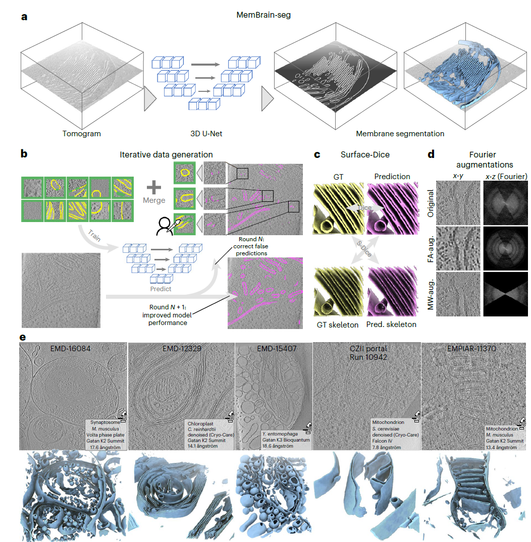
For deep learning models, what makes MemBrain v2 truly noteworthy extends beyond its architecture—it also lies in its approach to constructing training data. Rather than relying on single-source data, the research team adopted an iterative strategy akin to active learning. They continuously identified regions where their model struggled most and incorporated corrected versions of these “hard samples” back into the training set via manual refinement.
Initially, the project utilized patches derived from spinach (Spinacia oleracea) dataset annotations originally performed using TomoSegMemTV. Researchers then refined these patches manually using MITK Workbench. Each patch measured 160× voxels per dimension, assigning labels of background, membrane, or ignore independently for each voxel. This iterative refinement ultimately yielded 69 accurately annotated spinach chloroplast patches.
To enhance cross-platform and cross-imaging-condition generalizability, the methodology was subsequently extended to tomograms obtained from EMPIAR-11830 featuring Chlamydomonas reinhardtii. Due to differences between experimental setups, initial performance on this algal dataset lagged significantly behind results seen with spinach-derived inputs. To improve accuracy here, researchers iteratively relabeled 33 specific patches covering key ultrastructural features including thylakoid membranes, mitochondria, and Golgi apparatuses.
Furthermore, recognizing limitations imposed solely upon internal benchmarks, collaborators tested MemBrain-seg against independent proprietary collections while flagging areas requiring improvement before submitting corresponding new ground-truths after rigorous quality control checks resulting finally adding yet another batch comprising twenty-seven additional external validation sets contributing substantially towards increasing overall diversity present throughout entire corpus used during final stages development phase itself!
To further enhance model performance, the research team also leveraged existing publicly available membrane segmentation data sources. First, 40 membrane patches were generated using open-source tomogram simulators PolNet and CryoTomoSim, with each simulator producing 20 patches. Additionally, an externally annotated dataset was integrated by extracting 15 reliable membrane segmentation patches from the public DeePiCt dataset (EMPIAR-10988). All these regions were selected from areas where MemBrain-seg initially performed poorly, thereby further increasing the diversity of the training dataset.
MemBrain v2: An End-to-End Workflow for Membrane Analysis
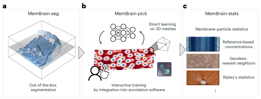
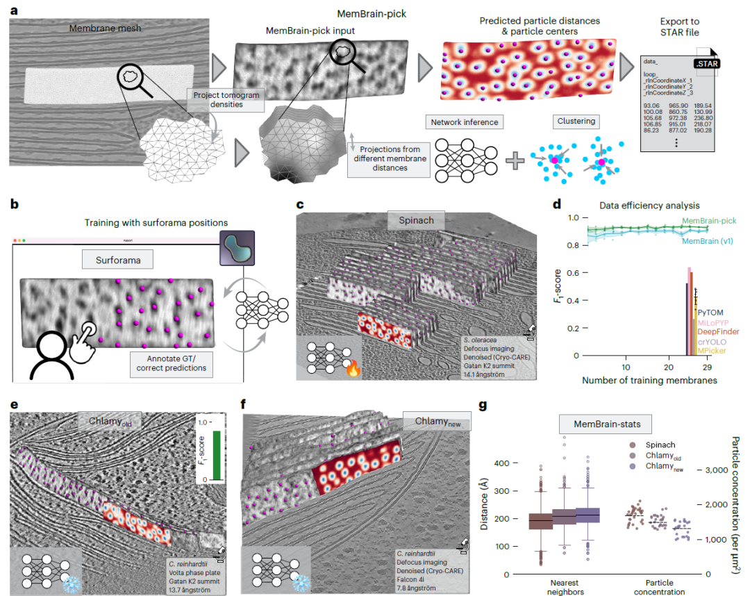
MemBrain v2 is a modular analysis workflow (shown below) designed to simplify the analysis of membranes and associated particles within cryo-ET datasets. Its three core modules—MemBrain-seg, MemBrain-pick, and MemBrain-stats—work synergistically to achieve membrane segmentation, particle localization, and quantitative analysis.
MemBrain-seg: A Generalized Method for Membrane Segmentation
MemBrain-seg (shown below) is a U-Net-based program that generates 3D membrane segmentation results from input tomograms via a single command line execution. This model was trained on a diverse and iteratively optimized dataset. The dataset was created through meticulous manual annotation and correction, developed in close collaboration with the research community to ensure coverage across various types and morphologies of membranes. Meanwhile, during network training, the research team introduced data augmentation methods tailored to the characteristics of cryo-electron tomography and loss functions specific to membranes. These strategies collectively ensured accurate and continuous delineation of membranes, facilitating subsequent visualization and downstream analyses.
MemBrain-pick: An Interactive Tool for Efficient Localization of Particle-Membrane Associations
MemBrain-pick (shown below) is specifically dedicated to efficiently localizing membrane-bound particles, such as membrane proteins. The research team trained neural networks to operate directly on membrane surfaces, enabling models to incorporate geometric information into predictions. This approach narrows the search space while improving both localization accuracy and data utilization efficiency. Furthermore, this module integrates with interactive tools based on Napari—such as Surforama—allowing researchers to quickly annotate and adjust particle positions, ensuring seamless transitions between ground truth (GT) data generation and model training.
MemBrain-stats: Quantitative Analysis of Particle Distribution
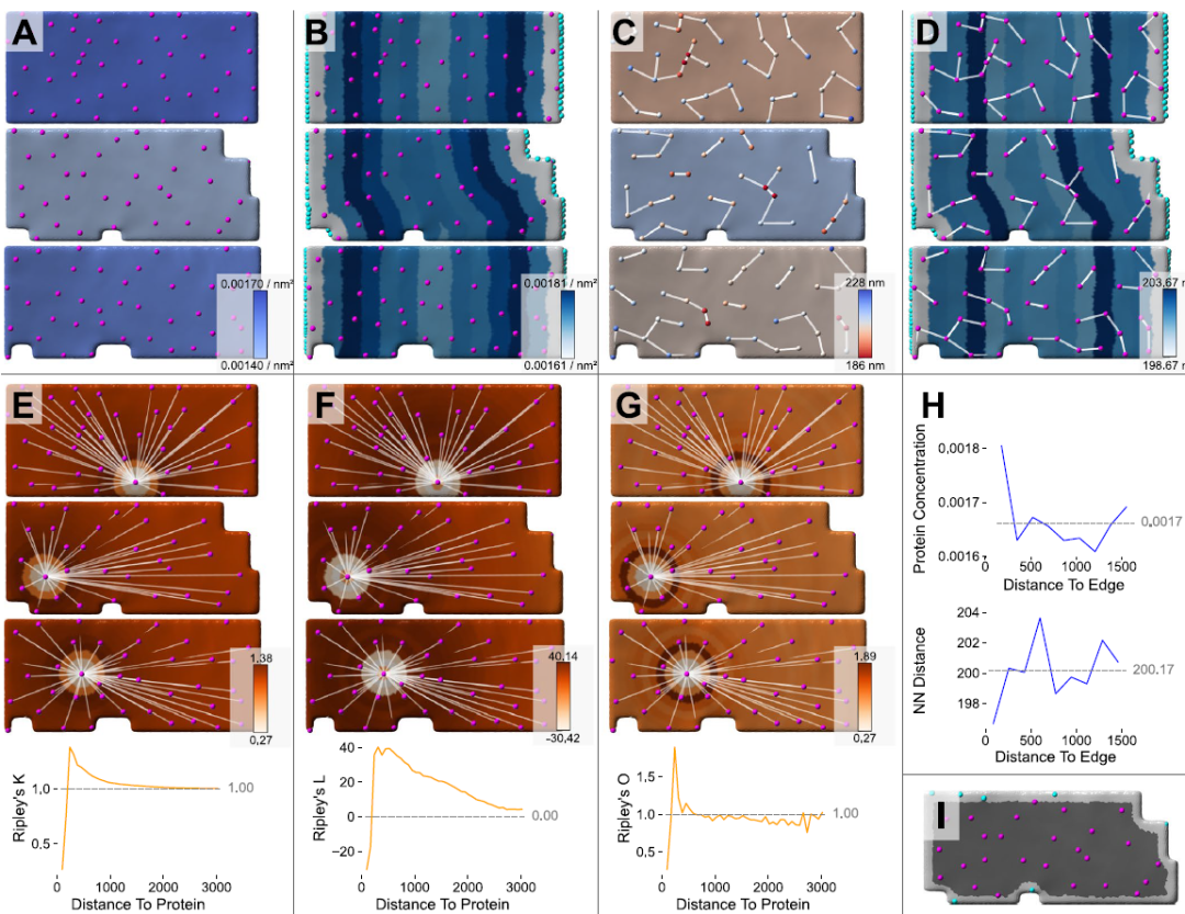
MemBrain-stats utilizes outputs from MemBrain-seg and MemBrain-pick to perform quantitative analyses of particle distributions on membrane surfaces. Key metrics calculated include particle density, geodesic nearest-neighbor distances, and Ripley's statistics (see figure below). By linking membrane morphology with patterns of particle organization, MemBrain-stats helps researchers investigate structural-functional relationships between membranes and their associated components more deeply within cryo-ET datasets.
Visualization Results of MemBrain-stats
Adopting a modular design, MemBrain v2 balances flexibility with ease-of-use throughout its framework. It offers simple yet powerful Command Line Interface (CLI), along with native integration support for Napari plugins to facilitate interactive annotations and visualizations.
Significant Improvements in Speed Throughput Achieved With MemBrain V2
Researchers utilized MemBrain stats to quantitatively analyze predicted thylakoid membrane protein distribution profiles comparing findings against previously published measurements covering all measured particles found stacked together inside chloroplasts shown here at Fig G . For spinach , obtained values closely matched those reported earlier regarding concentration levels per square micrometer area covered : there being approximately two thousand six hundred eighty eight molecules present versus one seven fourteen recorded before hand ; similarly chlamydomonas yielded slightly higher concentrations compared prior reports stating around fifteen ninety two units squared whereas newer estimates suggest closer proximity towards thirteen sixteen points zero five respectively indicating slight discrepancies possibly due methodological differences employed herein vis-a-vis previous studies conducted elsewhere globally speaking thus far concerning similar topics addressed therein accordingly !
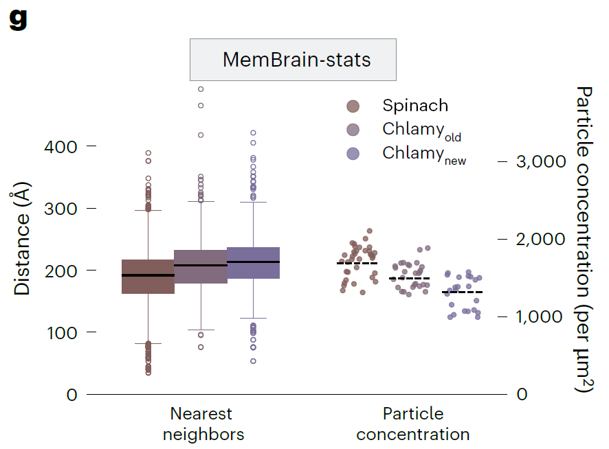
Overall, this streamlined MemBrain v2 workflow achieved analytical performance comparable to previous manual analyses while significantly improving processing speed and throughput for stacked thylakoid membrane studies. Researchers further utilized this pipeline to analyze higher-order organization patterns of other types of membrane-bound particles, including phycobilisomes and ribosomes.
Test Application: Organization of Phycobilisomes on Thylakoid Membranes
Researchers applied MemBrain v2 to a high-resolution cryo-electron tomography dataset of red algal chloroplasts (EMD-31244), demonstrating how the method efficiently extracts granule positions within phycobilisome chains and their spatial distribution patterns (see figure below).
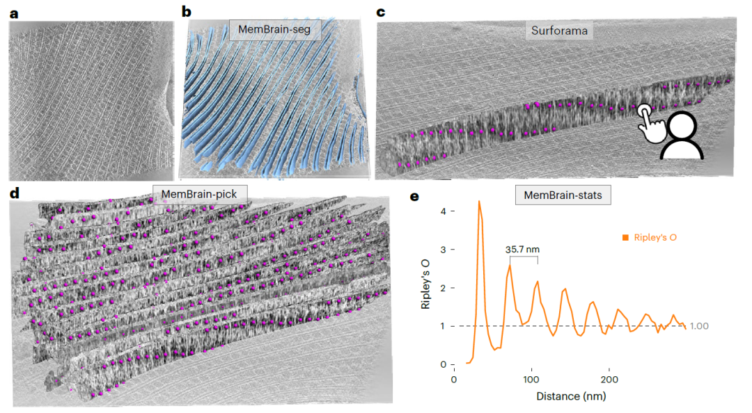
In the first step of processing, MemBrain-seg generated clearly separated segmentation results with high precision across all thylakoid membranes (Figure b above). Subsequently, researchers used the MemBrain lasso tool to perform connected component analysis, extracting individual membrane structural instances from the global segmentation results and visualizing them in Surforama. Leveraging this interactive visualization interface, researchers were able to manually annotate the locations of phycobilisome chain units among six selected membrane structures (Figure c above). These manually determined positions served as ground truth (GT) data to train the MemBrain-pick model. After training was completed, MemBrain-pick was applied to the remaining 23 membrane structures in the tilt series, successfully identifying phycobilisome locations throughout the entire volume. Prediction results revealed that phycobilisomes exhibit distinct periodic arrangements along the membrane surface (Figure d above).
To quantitatively analyze this spatial organization pattern, researchers further employed MemBrain-stats to calculate the Ripley’s K function. Results confirmed regular spacing between phycobilisomes, approximately 35 nm (Figure e above). This periodicity aligns closely with previously manually measured values of 34.5 nm, validating the reliability of the automated approach.
Test Application: Organization of Poly-ribosome Chains on Nuclear Envelopes
In another end-to-end application of MemBrain v2, researchers analyzed ribosome distributions on the outer nuclear envelope to identify localization and orientation patterns. When analyzing tomograms containing regions surrounding nuclei, MemBrain-seg accurately segmented both the nuclear envelopes and adjacent cytoplasmic membrane structures (Figure a below). Researchers then extracted specific nuclear envelope interests using the MemBrain lasso tool and visualized them in Surforama, enabling efficient annotation of GT ribosome positions (Figure b below).
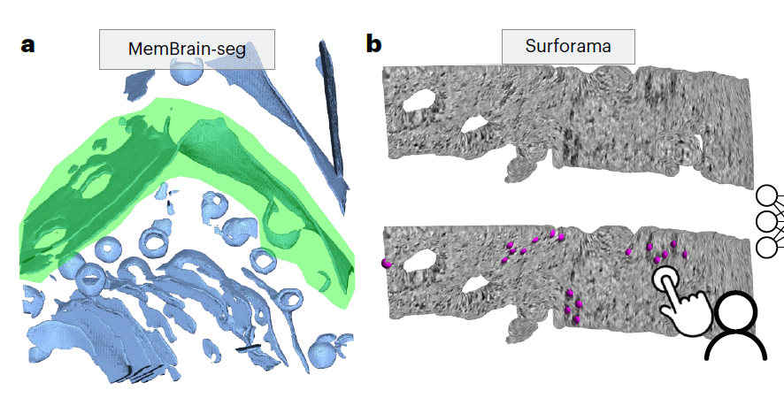
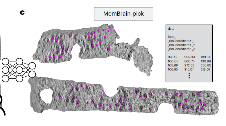
To automate ribosome localization, researchers initially annotated thirteen membrane structures to train the MemBrain-pick model; subsequently, they applied the trained model to predict positions in the remaining seventy-nine membrane structures, detecting a total of 4,515 ribosome sites (prediction examples see Figure c below).
To validate these predictions, researchers conducted subtomogram averaging (STA) using STOPGAP. The resulting structural map clearly presented an associated bound ribosome (Figure d below), which also included density signals corresponding to transmembrane TRAP complexes. Blurred peripheral densities appeared around the averaged structure, likely representing neighboring ribosomes—a finding corroborated by observing the spatial distribution of detected particles (Figure e below). Based on outputs provided by MemBrain-stats, researchers integrated constraints regarding angles, distances, and continuity between neighbors to extract poly-ribosome chains.
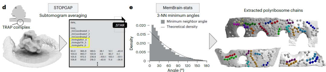
d, STA results. Left panel: Resulting average showing one associated bound ribosome.
e, Left panel: Histogram depicting minimum angle intervals among three nearest neighbors (3-NN).
This end-to-end MemBrain analysis process—starting from raw tomographic volumes through final identification of poly-ribosome chains—is expected to facilitate research into ribosomal biology across diverse organisms.
Conclusion
Certainly, MemBrain v2 does have its limitations. Its performance remains constrained by the diversity of training data, non-target membranous structures, and the inherent contrast and noise levels of cryo-ET itself. Accurate identification still poses challenges for weakly signaled or occluded particles, as well as small integral membrane proteins that can be easily obscured by strong membrane density. Moreover, segmentation thickness does not perfectly correspond to true biological membrane thickness.
In the future, expanding the training dataset further and integrating complementary tools such as TomoTwin could enhance its applicability across various membrane structures and protein complexes. More importantly, MemBrain v2 integrates membrane segmentation, particle localization, and spatial quantitative analysis into an open, user-friendly, and extensible workflow compatible with analytical frameworks including STAR, STA, and Surface Morphometrics. As more research teams participate in sharing data and tools, these AI-driven approaches hold promise for lowering the barrier to analyzing complex three-dimensional cryo-ET datasets, thereby advancing field practices away from manual-experience-dependent image recognition toward more automated, scalable, and reproducible quantitative analyses.
References:
https://phys.org/news/2026-09-ai-automates-3d-membrane-manual.html
https://phys.org/news/2026-09-ai-automates-3d-membrane-manual.html