Command Palette
Search for a command to run...
36 Million Cells Packed Into One Space, Stanford University Proposes Universal Cell Embedding Model UCE, Building a Massive cross-species Cell Atlas Across 8 Species
Over the past many years, cell biology has been dedicated to delineating the various phenotypes that cells can exhibit, the relationships between different states, and how cells transition between these states during development and disease.
The substantial growth in the scale of single-cell RNA sequencing (scRNA-seq) datasets has provided new opportunities to revisit such questions. However, existing computational methods still face difficulties in jointly analyzing these highly diverse datasets, because models may be affected by species-specific limitations, dataset-specific artifacts, or batch effects. Some computational methods for scRNA-seq data have been able to overcome certain limitations, but at the cost of: requiring model-specific tuning for each new dataset.
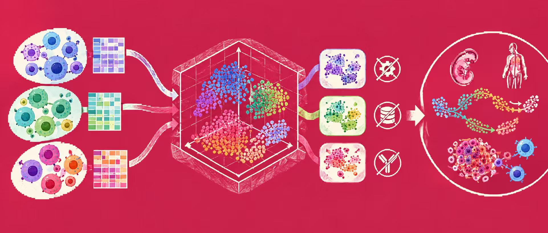
In this context, a research team from Stanford University proposed the universal cell embedding (UCE) foundation model. UCE possesses a unique capability: it generates representations for new single-cell gene expression datasets without the need for model fine-tuning or retraining, while maintaining robustness against dataset-specific and batch-specific artifacts. Furthermore, UCE neither requires cell type annotations nor preprocessing such as gene filtering of the input dataset. UCE can be applied to any set of protein-coding genes from any species, even if those genes have no homologous relationship with genes seen during model training. In this way, UCE learns a universal and intrinsically biologically meaningful representation of cell biology, enabling researchers to gain biological insights that go beyond what is directly observed in the experimental data.
The related research findings, titled "Universal cell embedding provides a foundation model for cell biology," have been published in Nature.
Research highlights:
-
For new cell data, without requiring data annotation, model retraining, or fine-tuning, UCE can map it into this unified representation space
-
The representations learned by UCE exhibit an emergent organizational structure of cell types and cell states, consistent with known biological principles
-
UCE can map new data into a universal embedding space, where annotated reference cell states already exist
Paper link:
https://www.nature.com/articles/s41586-026-10689-z
Building an IMA training dataset of over 36 million cells
The research team constructed a training dataset called the Integrated Mega-scale Atlas (IMA), integrating single-cell RNA sequencing data from various public sources. The core training data for UCE contains more than 36 million cells, of which the vast majority (33.9 million cells, 285 datasets) come from humans and mice, with the remainder consisting of 2.3 million cells from 28 datasets, covering 8 different species: human, mouse, zebrafish, rhesus macaque, cynomolgus monkey, mouse lemur, frog, and pig.
When visualizing the IMA, predicting green monkey cell types, matching cell type centroids of new species, and predicting Norn-like cells, the researchers did not use the full 36 million cells but instead used a representative sample of the IMA, to accelerate computationally intensive tasks such as UMAP calculations. This representative sample was constructed by randomly selecting 10,000 cells from each dataset without replacement. For datasets with fewer than 10,000 cells, the entire dataset was included directly. The resulting representative sample contained a total of 2,969,114 cells, with an average of 9,486 cells per dataset in the sample.
To evaluate the model's performance on truly "unseen" data, the paper also used multiple datasets that were not involved in model training. For example, Tabula Sapiens v.2 contains 581,430 cells, 27 tissues, 167 batches, and 162 distinct cell types, serving as an important zero-shot test dataset in the paper. The research team also prepared data from species not seen during training, such as green monkey, naked mole-rat, and chicken, to test whether UCE truly possesses cross-species generalization capability. Beyond these data, the paper further analyzed data such as mouse kidney and human lung disease, to verify whether UCE can discover new biological relationships from existing cell atlases.
All datasets analyzed in the paper are publicly available for download:
-
Green monkey lung and lymph node dataset: accession number GSE156755
-
Naked mole-rat dataset: accession number GSE132642
-
Chicken retina dataset: accession number GSE159107
-
Chicken heart dataset: accession number GSE149457
-
Mouse kidney dataset: accession number GSE193321
-
Human lung disease dataset: accession number GSE136831
UCE: A biology-informed cell foundation model
UCE overcomes the challenges of integrating scRNA-seq datasets by abstracting cells as "bags of RNA."
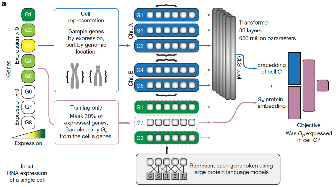
UCE is trained entirely in a self-supervised manner, without using any cell type annotations or dataset-based labels. As shown in the figure below, UCE first converts the RNA gene expression data of a single cell into an expression-weighted gene sample, and then uses a protein language model to represent the genes in the sample based on the proteins they encode. This enables UCE to meaningfully characterize any protein-coding gene from any species based solely on protein sequences, regardless of whether that species appears in the training data. After further integrating metadata such as gene chromosomal locations, this representation is fed into a large Transformer model. As a result, UCE can map cells from any tissue or species into a shared universal space without requiring additional training.
*Overview of the UCE model*
UCE's input consists of two parts: (1) scRNA-seq count data; (2) protein embeddings corresponding to the genes in the dataset, generated using the protein language model ESM2. The ESM2 protein language model takes amino acid sequences as input and outputs a numerical representation called a protein embedding*.
For a cell's gene expression count data, UCE weights and normalizes by expression level, and samples genes from that cell with replacement. This sample can only contain genes with non-zero expression, and the same gene can appear multiple times in the sample. These genes are then tokenized, i.e., converted into the protein embedding representations of the proteins they encode.
Genes belonging to the same chromosome are grouped by placing special markers and are sorted according to their genomic positions. Subsequently, a special marker representing the entire cell, the CLS token, is added to the very beginning of the cell representation. The combined representation is fed into the Transformer neural network, and the final embedding of the cell is taken from the embedding corresponding to the CLS token in the last layer of the Transformer.
Results: Gaining biological insights beyond what direct experimental observations can provide
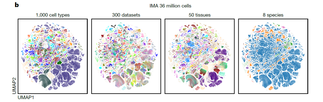
Based on UCE, the researchers further constructed an Integrated Mega-scale Atlas, embedding 36 million cells, encompassing more than 1,000 uniquely named cell types. Leveraging this unified space, they were able to further analyze how different cell types and tissues are organized within it.
UCE's embedding space exhibits emergent behavior
The study found that in the UCE space, cells naturally cluster based on biological conditions such as cell type, while cells from different experimental conditions, such as those from different batches, are able to mix with each other (panel b below). Since UCE only used unlabeled data during training, this organizational structure is not a result of the model being explicitly trained for it, but rather an emergent behavior of the model itself.
*UMAP visualization of the unified manifold in the UCE space*
The research team further investigated how tissue origin influences the state of different cell types. Although macrophages from different tissues exhibit diverse transcriptomic profiles, they remain highly aligned in the UCE space. For example, human macrophages are distributed across 73 different tissues, and for 72% (53) of these, the nearest centroid in the UCE space to a tissue-specific macrophage centroid is a macrophage centroid from another tissue.
Similar cross-tissue consistency can be observed in other abundantly present cell types, such as endothelial cells and neurons. This indicates that UCE, without explicit training or manual labels, can recognize the unique cell identity shared by macrophages across different tissues. This is an emergent organizational structure of UCE consistent with known biological principles, even though the model was not specifically trained for this phenomenon.
Zero-shot embedding of new datasets
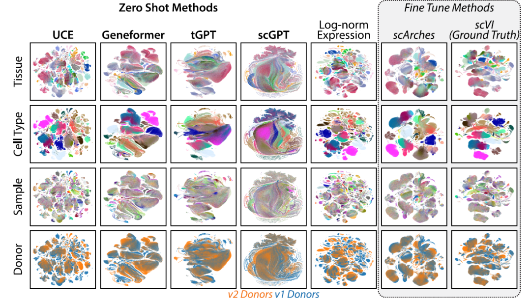
The researchers evaluated UCE's zero-shot performance on a new, unpublished dataset, Tabula Sapiens v.2. The results show that UCE's overall score is 13.9% higher than the best-performing Geneformer, with a 16.2% higher score for biological information preservation and a 10.1% higher score for batch effect correction. Compared to other methods, the embeddings generated by UCE can more clearly distinguish different cell types (as shown below).
*UCE outperforms existing methods in integrating Tabula Sapiens v2*
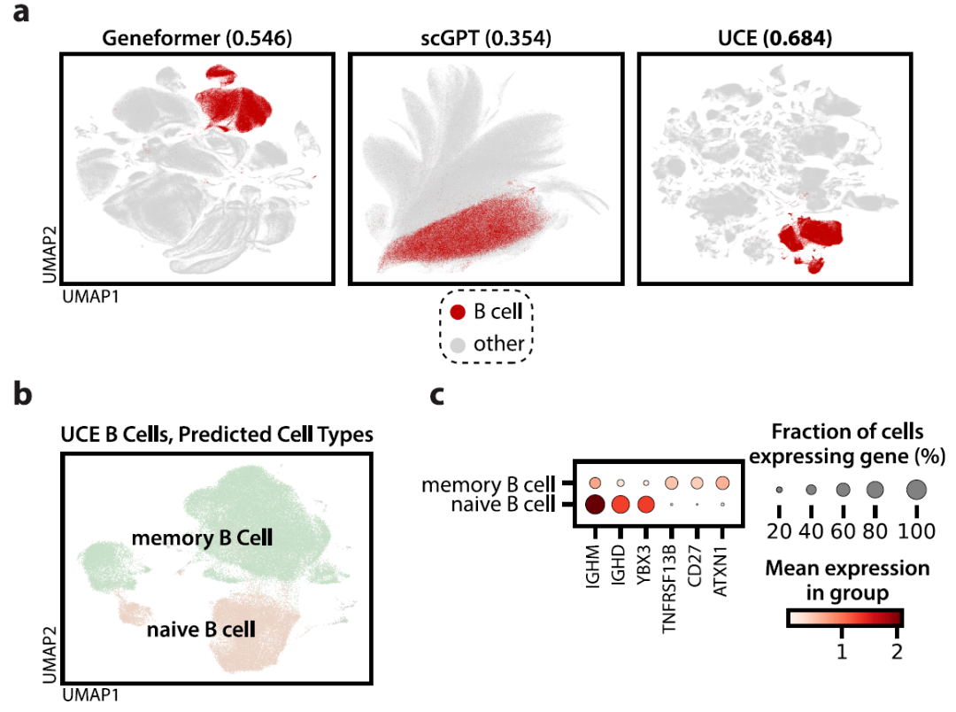
*UCE recaptures B cell identity in Tabula Sapiens v2*
Zero-shot embedding of new species
The researchers further compared UCE's performance with current state-of-the-art cross-species label transfer supervised methods, SATURN and SAMap. On 3 out of 4 datasets, UCE still outperformed SATURN and SAMap in transferring cell type labels between humans and new species. UCE was even able to generate consistent zero-shot embeddings for highly evolutionarily divergent species, such as fruit flies.
Organizational structure of cell types in new data
Beyond evaluation metrics focused on individual cell type clustering, the research team also examined the structure of the universal embedding space at a holistic level, analyzing the relative positions of different cells within this space. After embedding all cells from the Tabula Sapiens v.2 lung tissue, a cell type organizational structure with clear biological significance was observed (figure a below). Not only did different cell types cluster separately—such as T cells, monocytes, and endothelial cells forming distinct clusters—but higher-level cell categories, such as immune cells and epithelial cells, were also clearly distinguishable.
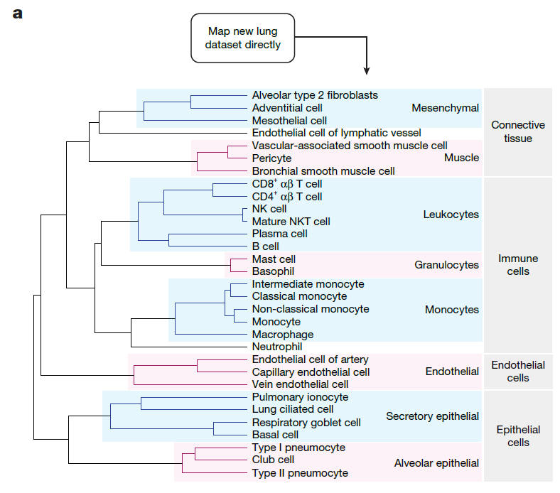
*The UCE space generated for new, previously unseen data exhibits a biologically meaningful organizational structure of cell types*
The researchers further compared this result with cell hierarchy structures derived from the Cell Ontology. The results showed that, compared with other zero-shot embedding methods, the cell clusters identified by UCE had higher similarity to the Cell Ontology.
Further experiments revealed that UCE can effectively learn a universal representation of cell biology. This representation not only distinguishes different cell types but also captures the relative similarities between cell types at different scales, potentially shedding light on deeper biological principles underlying cell development and function.
UCE analysis of lung disease outcomes
Finally, the researchers used UCE along with the previously constructed Norn cell classifier to study Norn-like cells in lung diseases. They sampled lung cells from patients with idiopathic pulmonary fibrosis (IPF), chronic obstructive pulmonary disease (COPD), and control patients, and generated their cell embedding spaces. In all three patient groups, Norn-like lung cells showing preferential expression of Norn marker genes were identified (figure d below); further analysis revealed that these Norn-like lung cells exhibited expression differences across disease groups (figure e below).
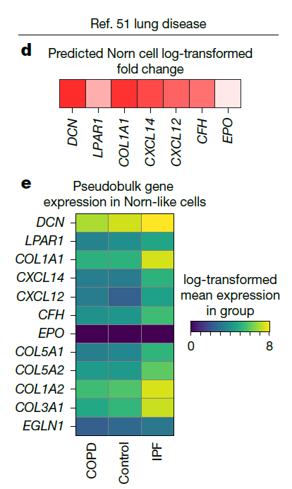
*The Norn cell case study demonstrates that UCE can support further analysis of single-cell datasets*
Both COPD and IPF are associated with elevated blood Epo levels, but Epo levels in COPD patients are higher than in IPF patients. Furthermore, compared with COPD patients, secondary erythrocytosis in IPF patients is less pronounced or occurs to a lesser degree. Given that Norn-like cells were found in lung tissue, and Norn cells can produce Epo, the differences in prognosis between these two diseases may be related to disease-associated differences in Norn-like cells.
Taken together, these results indicate that cells with transcriptional states similar to Norn cells can also be found in other tissues of the body, and these cells may play previously undescribed roles in disease processes. Because UCE is a universal model not restricted by tissue, species, or disease state, it can greatly facilitate the analysis of such large-scale, diverse data.
Conclusion
By constructing UCE, the researchers further expanded the ability to analyze scRNA-seq data. Although this model represents an important step toward universal cell embeddings, some limitations remain—current benchmarks primarily evaluate UCE's ability to recover expert-annotated cell types, but such tasks are inherently limited by the granularity and subjectivity of existing cell labels. Therefore, these evaluations may not fully reflect the model's ability to represent more nuanced biological processes, such as cellular responses to perturbations or integration across different modalities of data. Additionally, like other large-scale biological foundation models, UCE remains largely a black-box model, making it difficult to explain why and how certain embeddings form. Addressing this opacity will require further development of interpretability tools that connect the representations learned by the model to mechanistic biological understanding. Although UCE's training data is large in scale, it is still biased toward mammalian species, especially humans and mice, and also toward certain specific tissues, such as brain tissue, raising concerns about the model's ability to generalize to underrepresented species and biological scenarios.
Finally, it is worth emphasizing that although UCE can improve data alignment by capturing biological signals shared across datasets, it does not replace traditional batch effect correction methods. Traditional methods remain valuable for handling known experimental confounding factors.
References: